
Dr. Alan Detton, PhD
Assistant Professor in the Department of Pathology and Cell Biology at Columbia University Irving Medical Center
Dr. Detton courses in medical anatomy at the Columbia University Irving Medical Center (CUIMC). A foundational cornerstone of these courses was cadaver-based dissection, which required live, hands-on experiences. With the shift to remote teaching and learning in Spring 2020, Dr. Detton had to rethink how best to teach these anatomy courses, and how it would be possible to offer a dissection component that would allow students to have a traditional experience. Dr. Detton met the moment by leveraging available technology to reimagine learning opportunities for students. Read on to learn more about what Dr. Detton did in his course, what lessons and experiences he’s carrying forward, and the advice he has for other instructors at Columbia.
Leverage Available Technology to Reimagine Learning Opportunities for Students
Like other courses at VP&S, the anatomy lectures were delivered via Zoom to the entire class simultaneously. Students followed the power point-based material on their personal devices, responded to live quizzing, and submitted comments and questions much like they would do in person. Overall, after a small learning curve to the functionality of Zoom-based meetings the conversion of the didactic portion of the course was relatively straight-forward. What was less clear was an alternate solution to the cumulative 9 hours of weekly cadaveric-based dissection labs and 30-minute weekly review sessions for groups of approximately 20 students at a time. To compensate for the laboratory portion of the course I implemented two large educational innovations: synchronous zoom sessions with 3D anatomical models, and the creation of novel asynchronous materials.
Use 3D models to simulate in-person lab experiences.
Correlating to topics presented in the didactic lecture, eight 30-minute synchronous review sessions were given weekly to groups of approximately 28 students at a time. With the smaller group size, I was able to cover content from the previous week in a more intimate and informal setting, somewhat like the group laboratory review sessions previously provided. The sessions were highly interactive, not only due to the content being prescribed by student questions and requests, but as the resource utilized to teach the anatomical relationships was not still images in powerpoint but a 3D model-based app, Complete Anatomy.
The functionality in Complete Anatomy allows for digital anatomical structures to be individually selected, isolated, or hidden to create customized views of the human body which can be freely rotated or enlarged. Utilizing these features along with a “cut” tool, allowed me to mimic views of the human body typically encountered during dissection to reinforce key anatomical relationships often difficult to comprehend in static 2D images. Through live navigation of the 3D model, I was able to reinforce anatomical terminology and structural appearance with the students and provide highly visual repetition of content in a different manner from standard didactic lectures.
Though not a perfect replacement for the in-person laboratory sessions typically encountered by our students, student feedback of the synchronous 3D review sessions was the highest rated component of the course, demonstrating the power and potential of 3D models in anatomy courses and review sessions.
Since the pandemic, we have returned to dissection lab instruction, but Complete Anatomy is still provided as a supplement to the course and made available to the first-year students. Current research is transpiring looking at how to develop more accurate 3D models from cadaveric tissue to improve on the accuracy of the digital models representing the human form to compensate for limitations and inaccuracies within the platform. Inclusion of 3D model demonstrations in the main lectures has not yet occurred, but I feel this would be a powerful adaptation to traditional lectures due to the positive feedback from students on this manner of learning.
Create novel asynchronous course materials to engage students.
In addition to the synchronous didactic and review sessions, novel asynchronous material was provided to the students within Complete Anatomy and through posted cadaveric-based review videos. Utilizing the functionality in Complete Anatomy, I created custom models paired with interactive labels, images, and text, and organized them into blocks and lecture packets paralleling the organization of the gross anatomy course. In total, 3 blocks of material consisting of over 60 3D model-based lectures comprising approximately 615 interactive screens, recordings, and quizzes were provided for the students (See Figure A). By providing these resources through the platform, students could view the material when and where they chose as many times as needed or desired.
In addition to the digital 3D resources provided, I relied on my experience in having created over 78 cadaveric-based dissection videos, with over 13 hours of footage as part of the Grant’s Dissection Videos, which I authored along with the associated Grant’s Dissector text. Custom cadaver-based anatomy review videos of demonstrations on embalmed cadavers with labels and synced audio were created for our students (See Figure B). The videos focused on particularly difficult topics of the course including muscles of the forearm and hand, the contents of the axilla, innervation patterns of thoracic organs, and the peritoneum surrounding the abdominal organs. The videos were posted through a private YouTube channel, with links made available on CourseWorks.
In student feedback, many student comments related to the power and utility of the cadaveric videos, with students listing them as their preferred method of study and preparation along with many requests for an expanded library of videos. I have worked to expand the library of cadaveric-based videos, and hope to develop a more robust library of resources for future classes. Currently this expansion is taking place with the procedural anatomy correlates for second-year medical students preparing for their major clinical year with videos dedicated to anatomical structures related to spinal anesthesia, placement of chest tubes, and abdominal access for acute appendicitis and ectopic pregnancies.
Advice for Instructors and the Future of Teaching at Columbia
Be open to alternative modes of teaching and learning.
In Spring 2021, with some schedule adjustments, we were able to return to the dissection lab, and students were able to have some dissection experience working with teams composed of their fellow classmates, and perhaps most importantly, learn from donors who have generously given their remains for use in education. I still feel that cadaveric teaching is the best and most informative way to teach clinical gross anatomy as students get to feel real tissue, see examples of human variation, and learn to appreciate the complexity and beauty of the human form. But I also recognize that for our modern medical students, alternative methods of instruction also have a place. Students are adaptable – they value focused-resources intentionally crafted with learning goals in mind and tremendously enjoy non-traditional methods of content presentation. There is not only one way to teach anatomy.
Leverage students’ technological savvy to provide resources that resonate with them.
Leverage the technological savvy and learning styles of our amazing students to provide resources that resonate with them. While doing so we must ask what the major learning goals are, determine how this will be assessed, and then identify what resources can meet these needs. Moving forward as a community we need to provide quality learning opportunities both synchronously and asynchronously to be more inclusive to all our learners through equal access to high quality educational materials. We also need to carefully and intentionally pair technology with traditional teaching methods as appropriate, not be afraid of change, and embrace the opportunity challenges may present as we never know what may come out of that on the other side.
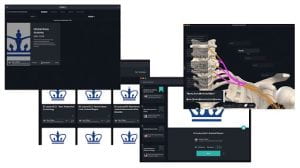
Figure A: Collage of screen captures of custom content in Complete Anatomy.
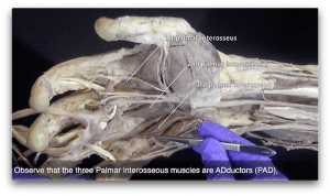
Figure B: Screenshot of video for the superficial dissection of the palm of the hand.